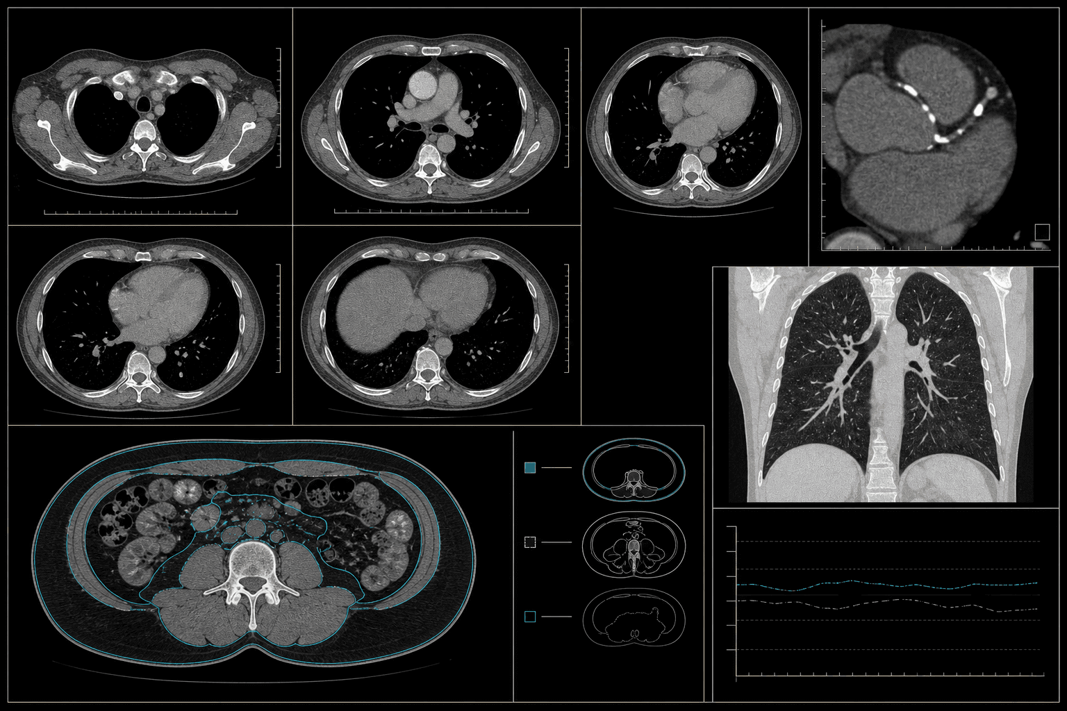
One scan can contain multiple signals
A non-contrast chest CT may support coronary calcium, cardiac chamber, lung, vertebral bone or body-composition analysis. That does not mean one cohort automatically validates every application. Field of view, reconstruction kernel, slice thickness, dose and reference measurements must match the intended task.
The CT programme control plane
A useful request names the anatomical coverage, acquisition constraints, outcome or measurement and intended validation design. It also states whether historical routine-care scans are acceptable or whether a controlled protocol is required.
- Scanner vendor, model, tube settings and reconstruction kernel
- Slice thickness, spacing, field of view and dose information
- Reference measurements, reports, outcomes or expert labels
- Positive, negative and difficult-case composition
- Hospital, scanner and demographic distributions for holdout testing
Validation is a separate design problem
A development cohort and an external validation cohort should not share patients or leak repeated examinations across splits. Site and scanner independence, prevalence and demographic representation are documented so performance can be interpreted correctly.
Questions, answered directly.
Is contrast-enhanced CT included?+
This programme is currently framed around non-contrast CT. Contrast-enhanced studies would require a separate specification.
Can the same CT support several labels?+
Potentially, but each label needs its own ground-truth method and acceptance criteria.
Is this a ready-to-download dataset?+
No. Access is controlled, and each release is subject to hospital authority, privacy, programme composition and licensing controls.